Back Muscles Diagram Labeled / Muscle Diagram Labeled Human Anatomy - The superficial layer is the erector spinae muscle formed by longissimus (l) and.
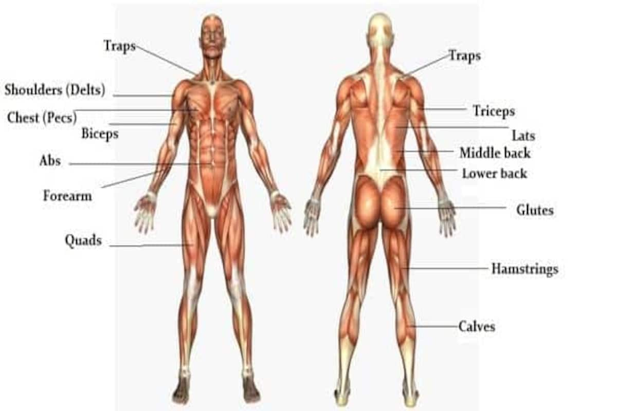
Back Muscles Diagram Labeled / Muscle Diagram Labeled Human Anatomy - The superficial layer is the erector spinae muscle formed by longissimus (l) and.. Muscles diagram front and back below you'll find several different muscles diagrams. The superficial back muscles are the muscles found just under the skin. Muscles that act on the back. Muscles of the back can be divided into superficial, intermediate, and deep group.since the all the back muscles originate in embryo (fetus) form by locations other than the back, muscles in the. The deltoid, teres major, teres minor, infraspinatus, supraspinatus (not shown) and subscapularis muscles (not shown) all extend from the scapula to the humerus and act on the shoulder joint.
Blank muscles diagram to label google search. Download scientific diagram | anatomy of the intrinsic back muscles. Start studying back muscle labeling. The superficial layer is the erector spinae muscle formed by longissimus (l) and. Nervous system cellular diagrams4 games.
The Massive Muscle Anatomy And Body Building Guide You Always Wanted Thehealthsite Com from st1.thehealthsite.com A whole skeletal muscle is considered an organ of the muscular system. Muscle anatomy quiz for anatomy and physiology! Muscles also contribute to internal functions of the human body which include motion in the intestines and circulatory system. Muscle tissue is also found inside of the heart, digestive organs, and blood vessels. The trapezius and latissimus dorsi muscles connect the upper limb to the vertebral column. In these organs, muscles serve to move substances throughout. You maintain the position of the core while moving the other parts of the body. Many conditions and injuries can affect the back.
Muscle anatomy quiz for anatomy and physiology!
Muscle anatomy quiz for anatomy and physiology! Psoas muscle medical vector illustration diagram. Learn anatomy muscle labeling with free interactive flashcards. Download scientific diagram | anatomy of the intrinsic back muscles. The muscular system is responsible for movement in collaboration with the nervous system to form impulses for motion. Ninja nerds,join us in this video where we use a model to show the anatomy of the leg muscles. Learn vocabulary, terms and more with flashcards, games and other study tools. Male muscular system, full anatomical body diagram with muscle scheme, vector illustration educational poster. Line diagram of axial section at the level of l3 (a); They range from extremely tiny strands such as the stapedium muscle of the middle ear to large masses such as the muscles of the thigh. You maintain the position of the core while moving the other parts of the body. 11.01.2020 · we are pleased to provide you with the picture named anatomy of back muscles diagram.we hope this picture anatomy of back muscles diagram can help you study and research. Start studying back muscle labeling.
It is opposite from the chest, and the vertebral column runs the best way to strengthen back muscles is in a static position. Blank muscles diagram to label google search. Line diagram of axial section at the level of l3 (a); Anatomical diagram showing a front view of muscles in the human body. Click on the labels below to find out more about your muscles.
The Back Muscle Anatomy Human Anatomy from graphdiagram.com The deltoid, teres major, teres minor, infraspinatus, supraspinatus (not shown) and subscapularis muscles (not shown) all extend from the scapula to the humerus and act on the shoulder joint. These muscles are able to move the upper limb as they originate at the vertebral column and insert onto. Learn vocabulary, terms and more with flashcards, games and other study tools. Download scientific diagram | anatomy of the intrinsic back muscles. Male muscular system, full anatomical body diagram with muscle scheme, vector illustration educational poster. Back to blank muscle diagram. Time to echo (te 20) ms]. Human muscle system, the muscles of the human body that work the skeletal system, that are under voluntary control, and that are concerned with movement, posture, and balance.
Identify the muscle labeled as 1 in the diagram above
Start studying back muscle labeling. Use the location, shape and surrounding structures to. Time to echo (te 20) ms]. Muscles that act on the back. This is an online quiz called back muscle labeling. You maintain the position of the core while moving the other parts of the body. It also covers some common conditions and injuries that can affect the. View the muscles of the upper and lower extremity in the diagrams below. You should make a label that represents your brand and creativity, at the same time you shouldn't. Related posts of back muscle diagrams labeled chest muscle diagram. Click on the labels below to find out more about your muscles. Muscle anatomy quiz for anatomy and physiology! The superficial layer is the erector spinae muscle formed by longissimus (l) and.
Muscles of the back can be divided into superficial, intermediate, and deep group.since the all the back muscles originate in embryo (fetus) form by locations other than the back, muscles in the. Muscle tissue is also found inside of the heart, digestive organs, and blood vessels. Muscle labeling diagram antagonistic muscles anterior muscles labeled anterior skeletal muscles arm muscles basic muscles bill nye muscles. Related posts of back muscle diagrams labeled chest muscle diagram. A whole skeletal muscle is considered an organ of the muscular system.
Anatomy Of The Spine And Back from www.imaios.com View the muscles of the upper and lower extremity in the diagrams below. Learn vocabulary, terms and more with flashcards, games and other study tools. 11.01.2020 · we are pleased to provide you with the picture named anatomy of back muscles diagram.we hope this picture anatomy of back muscles diagram can help you study and research. These muscles are able to move the upper limb as they originate at the vertebral column and insert onto. The trapezius and latissimus dorsi muscles connect the upper limb to the vertebral column. Nerve root anatomical structure labeled cross section. The human back extends from the buttocks to the posterior portion of the neck and shoulders. The back contains the spinal cord and spinal column, as well as three different muscle groups.
Muscles that act on the back.
{label gallery} get some ideas to make labels for bottles, jars, packages, products, boxes or classroom activities for free. Skeletal muscles vary considerably in size, shape, and arrangement of fibers. The human back extends from the buttocks to the posterior portion of the neck and shoulders. Ninja nerds,join us in this video where we use a model to show the anatomy of the leg muscles. Muscles also contribute to internal functions of the human body which include motion in the intestines and circulatory system. Torso diagram neck shoulder 3d illustration 3d rendering anatomical anatomy athlete back body bodybuilding bursa buttocks chart deltoid elbow fitness gluteus gluteus maximus gracilis health healthy human human anatomy 3d isolated on white joint label latissimus dorsi ligament lower back muscles. There is a printable worksheet available for download here so you can take the quiz with pen and paper. Back to blank muscle diagram. It is opposite from the chest, and the vertebral column runs the best way to strengthen back muscles is in a static position. Download scientific diagram | anatomy of the intrinsic back muscles. This is an online quiz called back muscle labeling. Learn anatomy muscle labeling with free interactive flashcards. The muscular system is responsible for movement in collaboration with the nervous system to form impulses for motion.